티스토리 뷰
측면발목인대 해부학
Posterior talofibular ligament, Anterior talofibular ligament, Calcaneofibular ligament
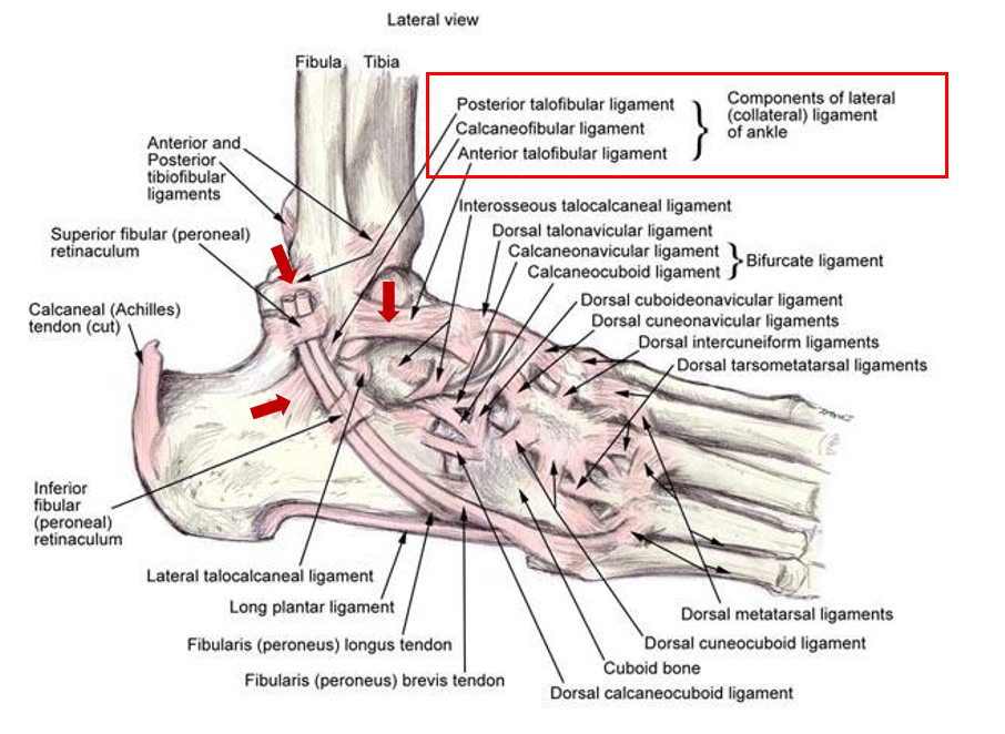
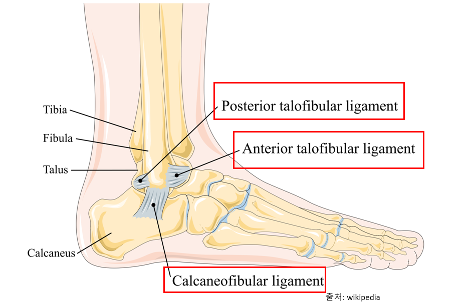
[Lateral ligament of ankle]
기능: 과도한 발목 내번 제한
손상 기전: 과도한 발목의 내번
발목이 꺾이면 손상되는 대표적인 인대 3가지로, 발목이 과도하게 꺾이게 되면 무조건 이 중 하나 이상이 손상된다.
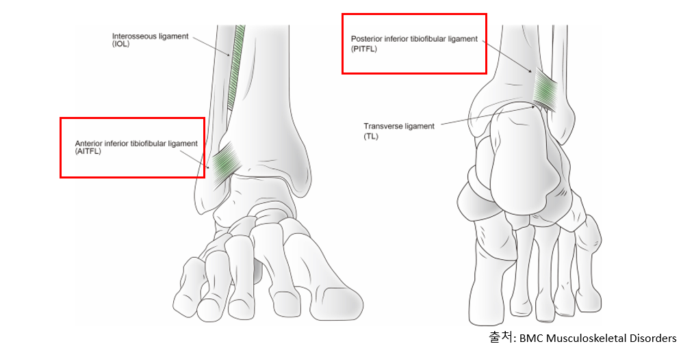
Anterior and Posterior tibiofibular ligament
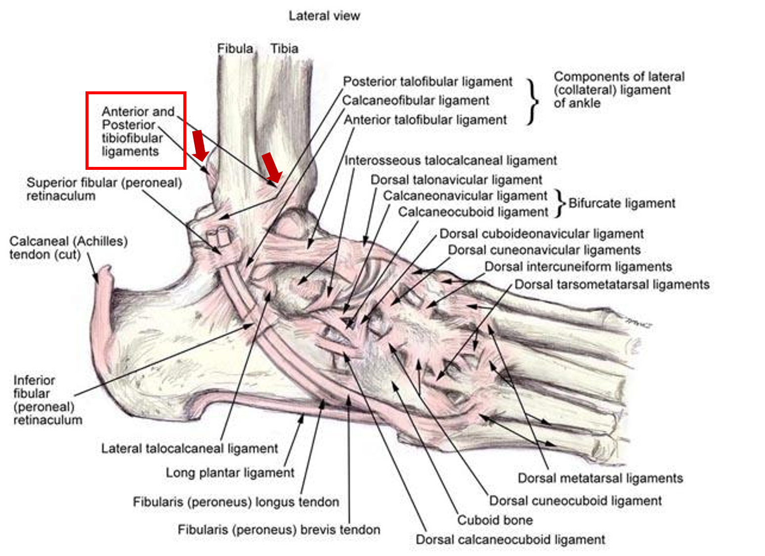
기능: 과도한 비골 외회전, 발목 내번 제한
손상 기전: 발목 골절 및 과도한 내번
해당 인대에 손상이 발생하면,
발목을 내번 하거나 발을 외회전 하는 것에 제한이 생길 가능성이 높다.
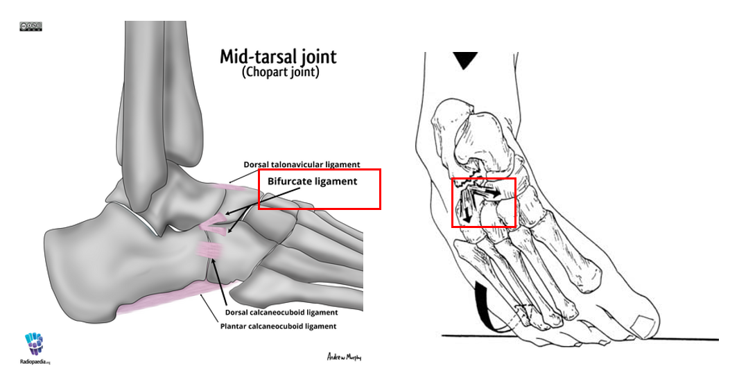
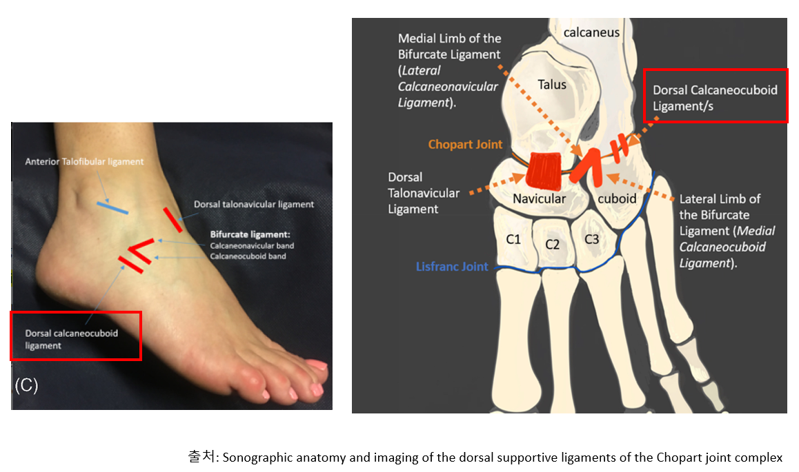
Bifurcate ligament
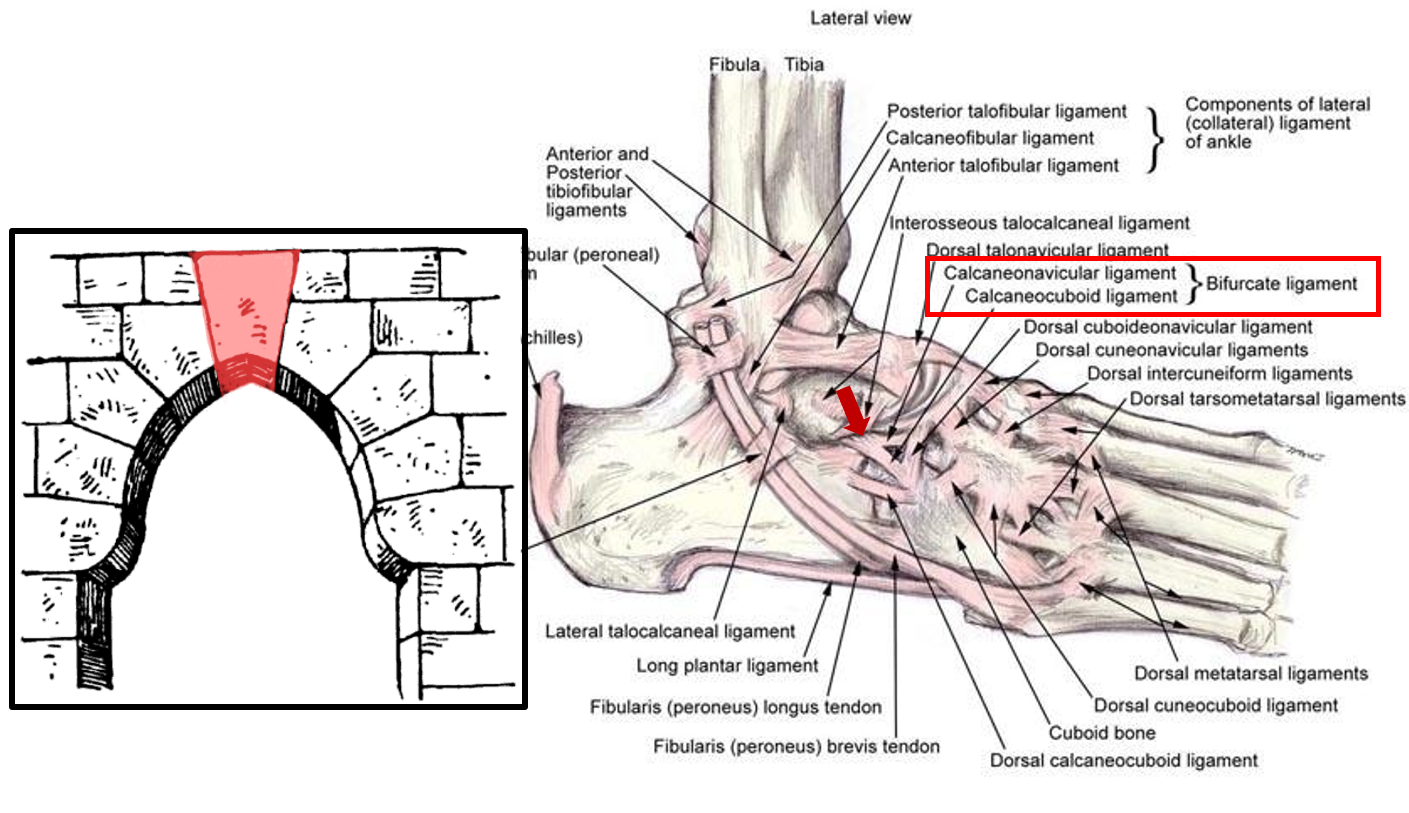
기능: Calcaneal을 navicular, cuboid에 단단히 고정시켜 발목의 안정성의 key stone으로 불림
손상 기전: 발목 골절 및 과도한 내번
Y모양의 강력한 인대로, 손상되면 아치의 안정성이 무너져서
달리기 할 때 발에 제대로 힘을 주기 어려움
Interosseous talocalcaneal ligament
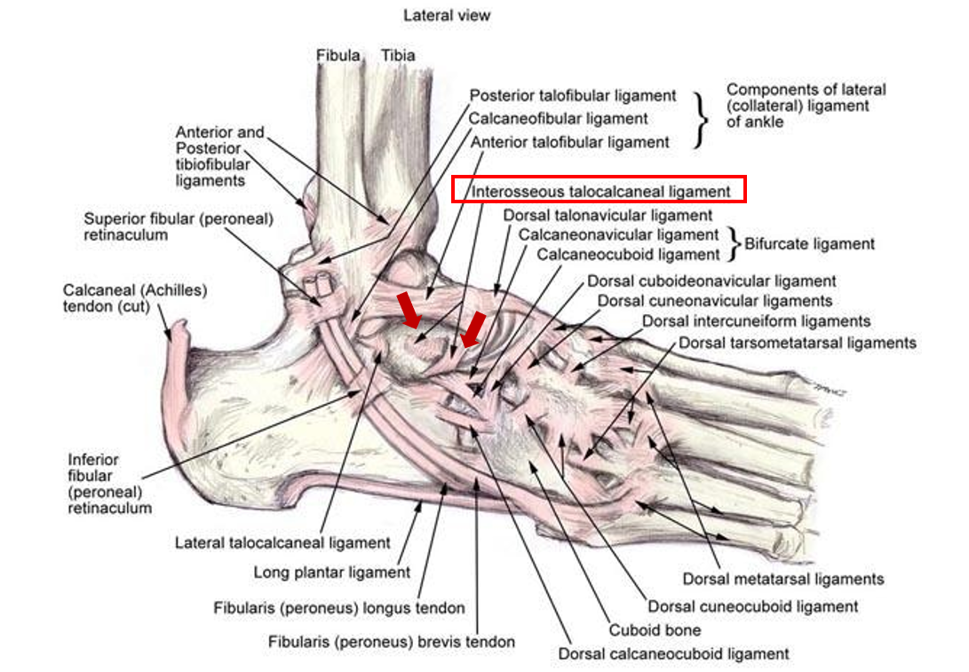
기능: talus를 calcaneal에 고정시켜주는 인대로, 거골하관절의 과도한 움직임을 제한함
손상 기전: 발목 골절 및 과도한 내번
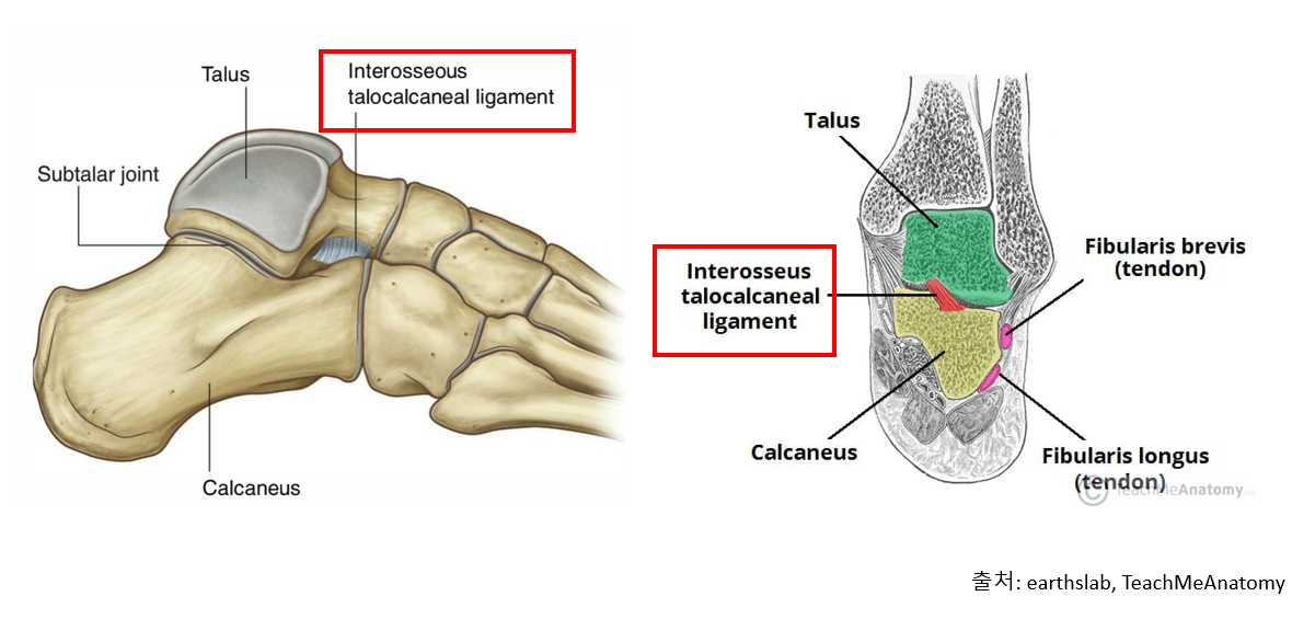
거골하관절의 과도한 회내를 제한하며, 손상되는 경우,
발의 아치가 과도하게 무너져서 발의 충격이 증가할 수 있음 거골하관절 안정성의 핵심 중 핵심
Dorsal talonavicular ligament
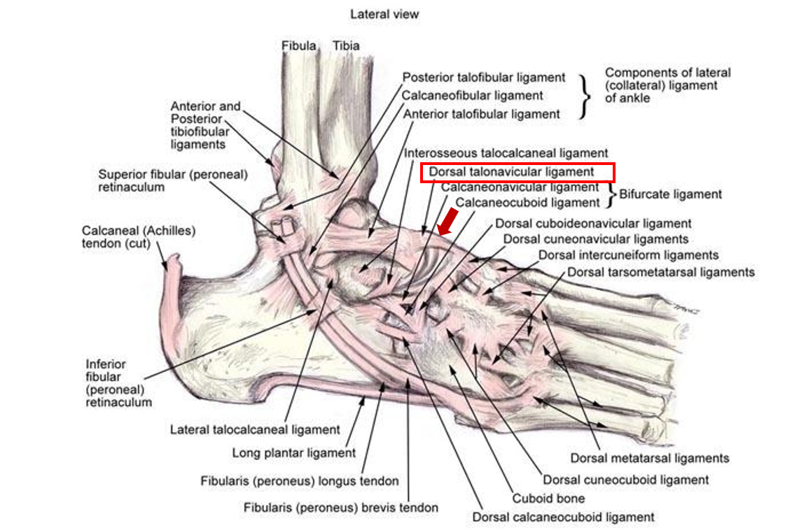
기능: talus와 navicular를 연결해주는 인대로, 발목의 저측굴곡을 제한
손상 기전: 저축굴곡을 동반한 내번
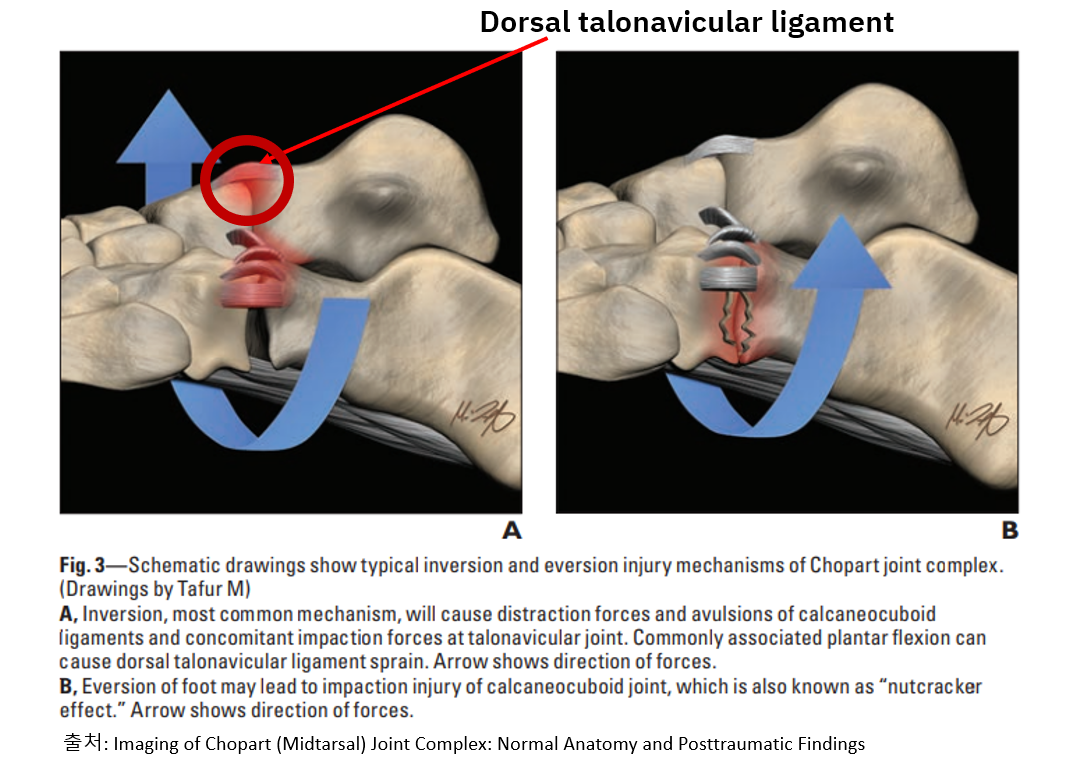
De Dea et al에 의하면, 발목 염좌의 22%는 이 인대의 손상이 동반됨.
발목이 꺾일 때 저측굴곡이 동반되는 경우가 많은 걸 생각해보면 당연한 사실
Dorsal calcaneocuboid ligament
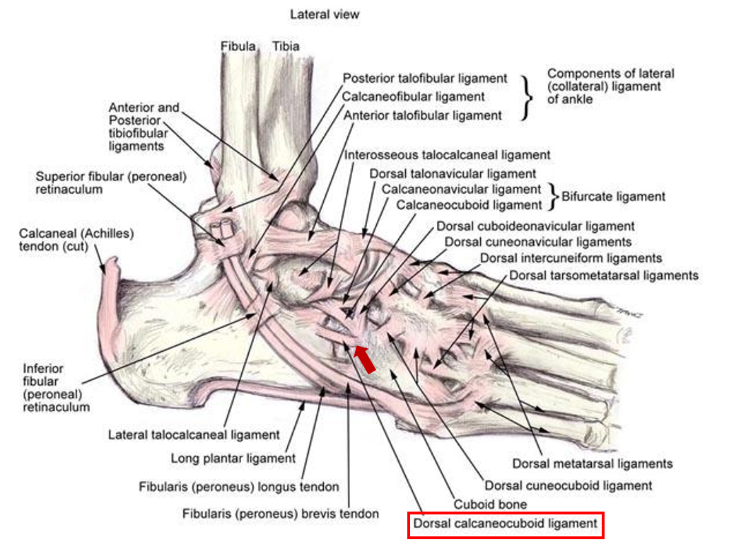
기능: calcaneofibular ligament와 유사한 기능
손상 기전: 발목의 과도한 내번
발목 내번 부상 시 30%는 해당 인대가 손상되며,
발목 바깥쪽 불안정성 및 통증의 주범이 되기도 함
Dorsal cuneocuboid, cuboideonavicular, cuneonavicular ligament
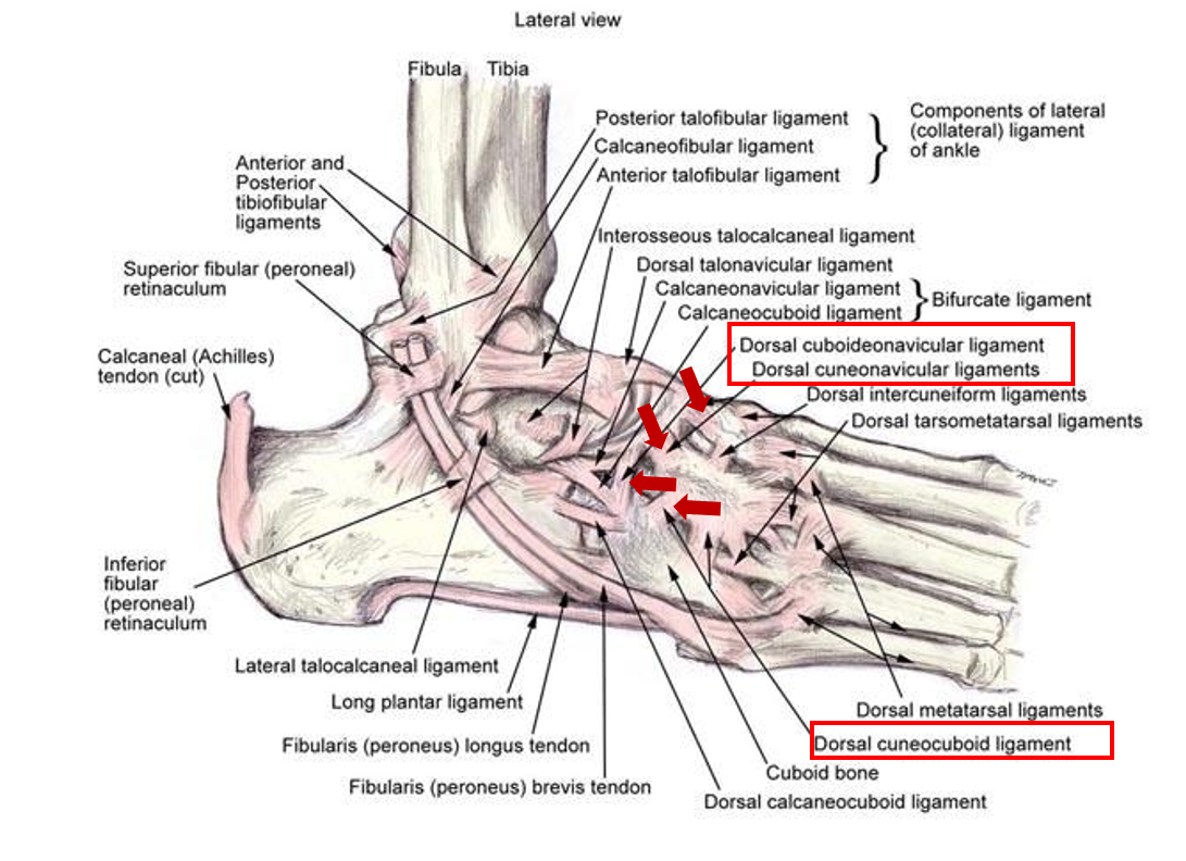
기능: midfoot의 안정성을 제공하는 인대들로, 아치의 안정성을 유지해줌
손상 기전: 발목 골절, 스포츠 손상
발목 안정성이 극대화되는 저측굴곡 상태에서 외부의 충격이 가해지는 경우,
교통사고 등으로 인해 손상됨
Lateral talocalcaneal ligament
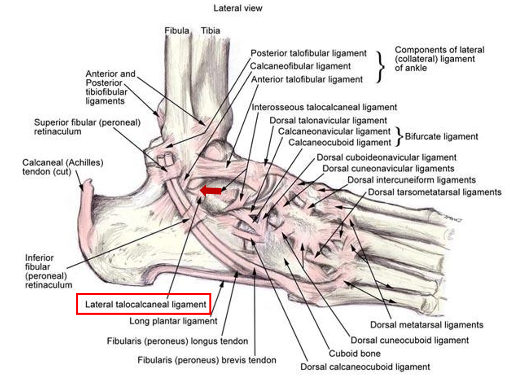
기능: calcaneofibular ligament와 유사한 기능
손상 기전: 발목의 과도한 내번
참고
Carto, C., Lezak, B., & Varacallo, M. (2019). Anatomy, Bony Pelvis and Lower Limb, Distal Tibiofibular Joint (Tibiofibular Syndesmosis).
Kafka, R. M., Aveytua, I. L., Choi, P. J., DiLandro, A. C., Tubbs, R. S., Loukas, M., ... & D'Antoni, A. V. (2019). Anatomico-radiological study of the bifurcate ligament of the foot with clinical significance. Cureus, 11(1).
Poonja, A. J., Hirano, M., Khakimov, D., Ojumah, N., Tubbs, R. S., Loukas, M., ... & D'Antoni, A. V. (2017). Anatomical study of the cervical and interosseous talocalcaneal ligaments of the foot with surgical relevance. Cureus, 9(6).
Tochigi, Y., Amendola, A., Rudert, M. J., Baer, T. E., Brown, T. D., Hillis, S. L., & Saltzman, C. L. (2004). The role of the interosseous talocalcaneal ligament in subtalar joint stability. Foot & ankle international, 25(8), 588-596.
De Dea, M., L Loizou, C., Allen, G. M., Wilson, D. J., Athanasou, N., Uchihara, Y., ... & Cosker, T. (2017). Talonavicular ligament: prevalence of injury in ankle sprains, histological analysis and hypothesis of its biomechanical function. The British journal of radiology, 90(1071), 20160816.
Tafur, M., Rosenberg, Z. S., & Bencardino, J. T. (2017). MR imaging of the midfoot including Chopart and Lisfranc joint complexes. Magnetic Resonance Imaging Clinics, 25(1), 95-125.
Walter, W. R., Hirschmann, A., Tafur, M., & Rosenberg, Z. S. (2018). Imaging of Chopart (midtarsal) joint complex: normal anatomy and posttraumatic findings. American Journal of Roentgenology, 211(2), 416-425.
Fenech, M., & Wylie, B. (2022). Sonographic anatomy and imaging of the dorsal supportive ligaments of the Chopart joint complex. Sonography, 9(2), 83-91.
Angin, S., & Simsek, I. (Eds.). (2020). Comparative kinesiology of the human body: normal and pathological conditions. Academic Press.
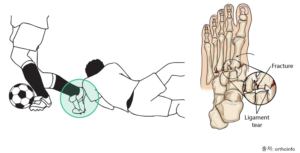
https://orthoinfo.aaos.org/en/diseases--conditions/lisfranc-midfoot-injury
'Exercise is medicine > 기능 해부학' 카테고리의 다른 글
| 상완신경총(The Brachial Plexus) (0) | 2023.09.21 |
|---|---|
| 요천추신경총 해부학 (0) | 2018.03.19 |
| 견갑대 해부학 (2) | 2018.03.16 |
| 골반 해부학 (1) | 2018.03.15 |
| 목 뼈 해부학 (0) | 2018.03.12 |


- Total
- Today
- Yesterday