티스토리 뷰
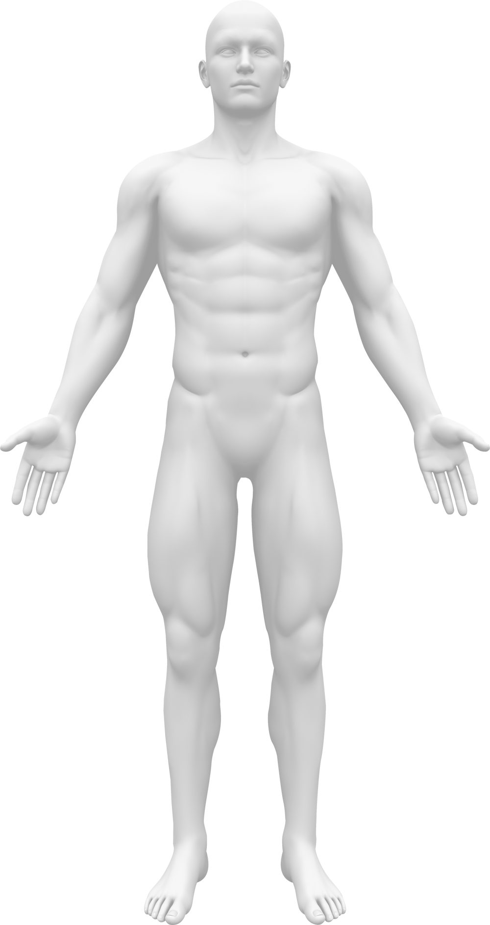
상완신경총
우리 몸에는 경신경총, 상완신경총, 요천추신경총 등 다양한 신경총이 존재하는데, 이 중 상완신경총은 C5~T1에서 나온 경추신경이 모여 이루어지는 신경다발을 의미한다.
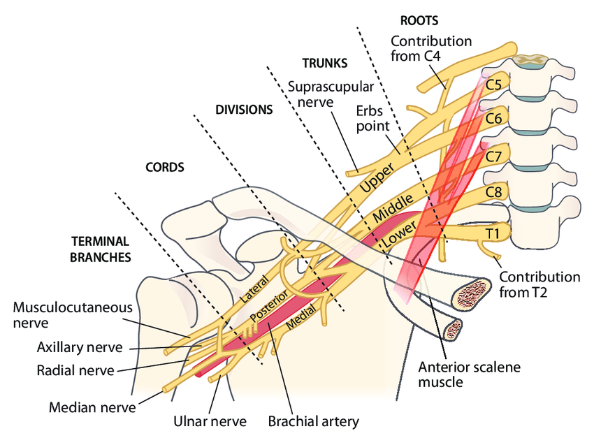
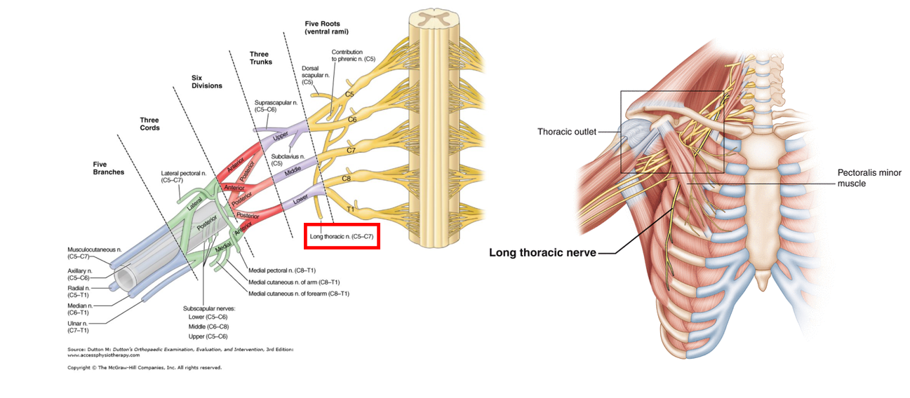
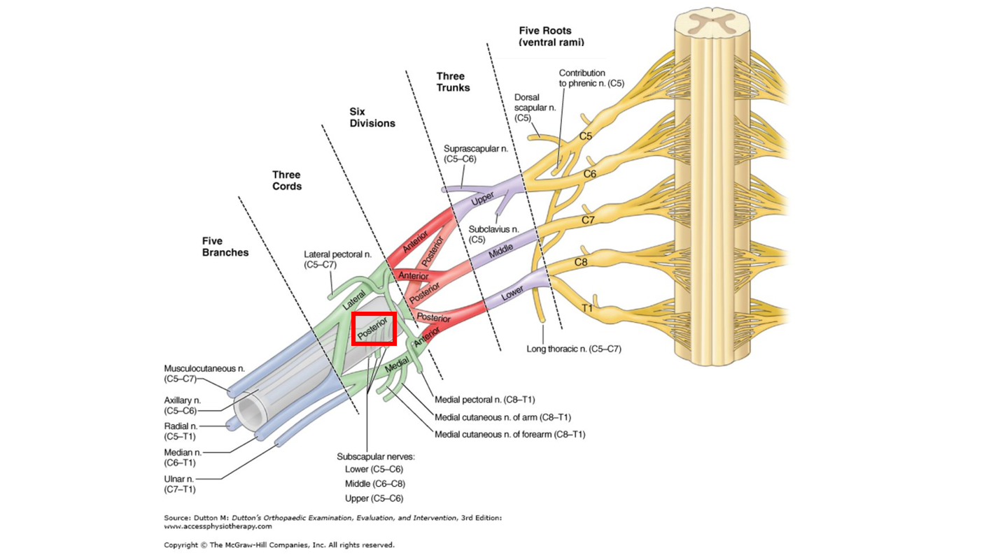
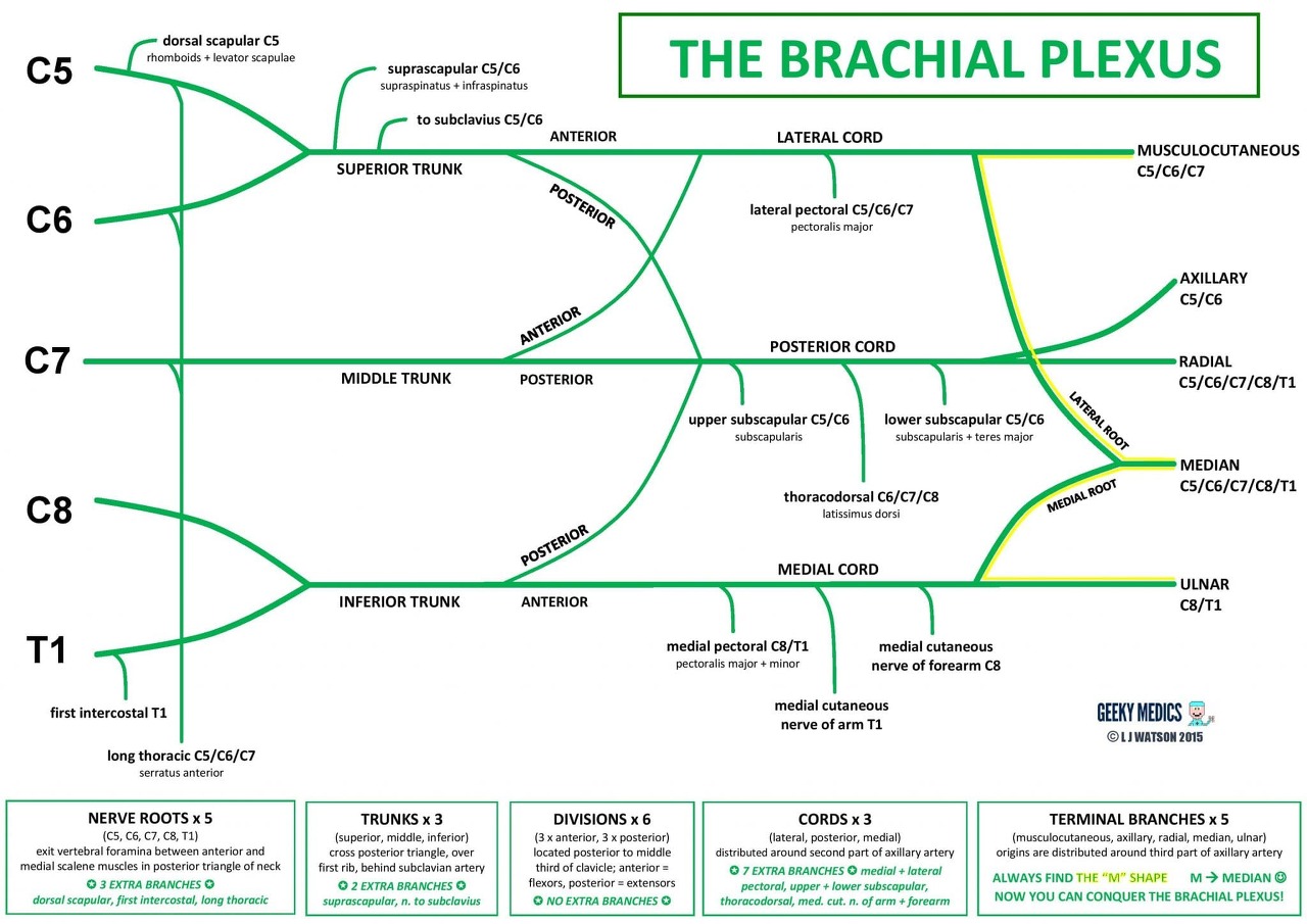
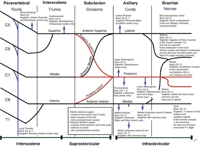
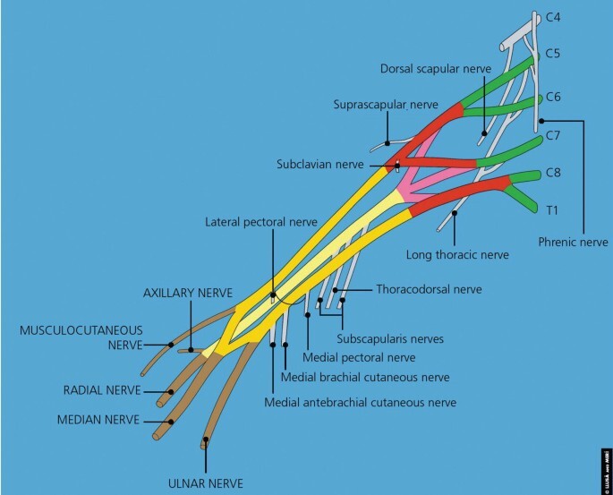
상완신경총은 5개의 신경근(Root), 3개의 신경 간부(Trunk), 6개의 신경 분지(Division), 3개의 신경 신경속(Cord), 5개의 신경말단 분지(Terminal branch)로 나뉜다.
신경근(Root)
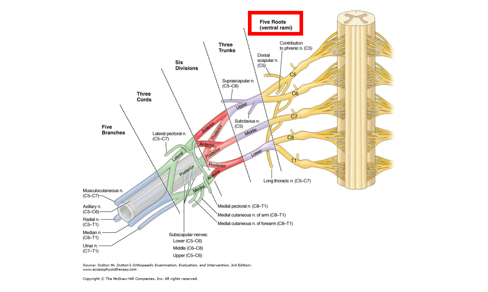
신경근(Root)은 C5~T1 척수에서 나온 경추신경 중, 전방가지(Ventral ramus)의 모음을 의미하며, 총 5개의 신경근이 존재한다.
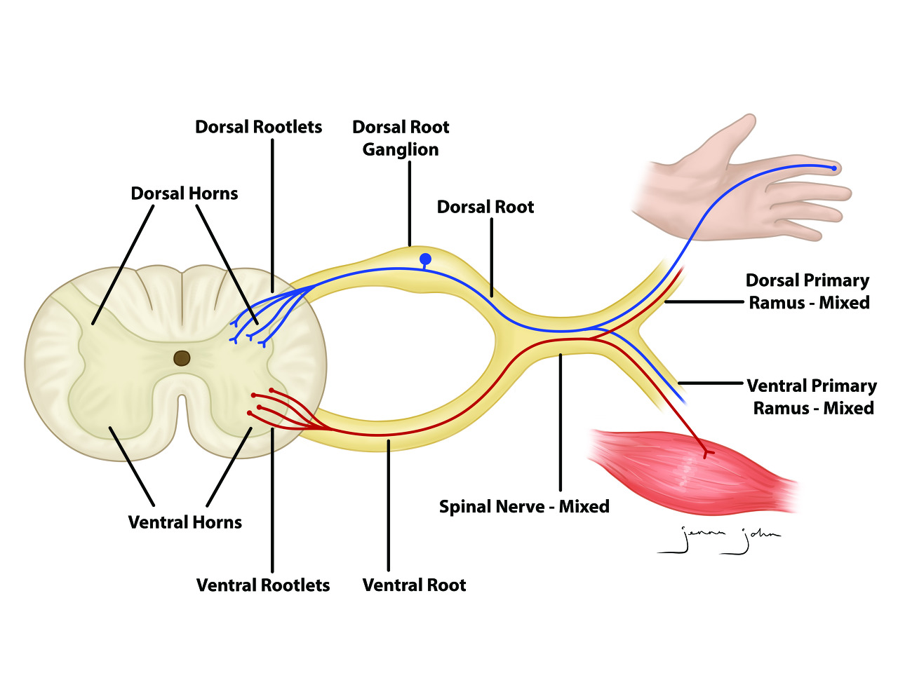
척수 신경은 위 그림에서 보이는 것처럼 감각 정보를 담당하는 Dorsal root와, 운동 정보를 담당하는 Ventral root로 나뉘는데, 이렇게 분지된 신경들은 다시 합쳐져서 Dorsal ramus(등쪽 가지)와 Ventral ramus(배측 가지)로 분지된다.
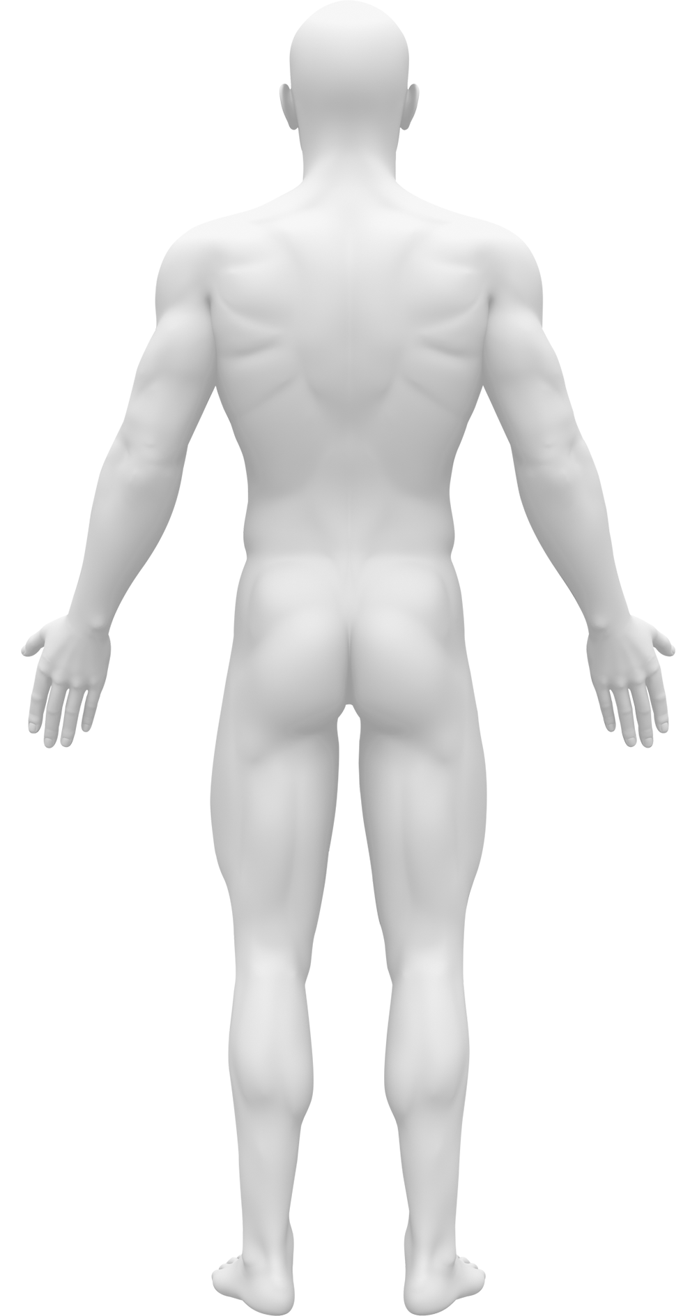
이 중 Dorsal ramus는 몸 뒤쪽 피부와 심부 근육들을 지배하고, Ventral ramus는 몸 앞쪽, 외측, 상지 그리고 하지를 지배한다.
신경근 손상
Roots가 압박되는 경우 각 신경근이 담당하는 피부 분절과 근육 분절에 문제가 발생한다.
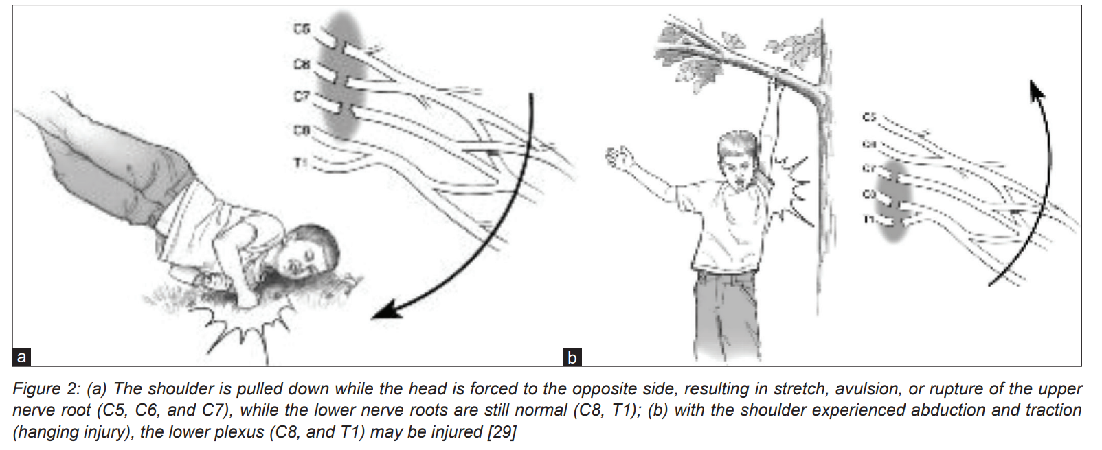
신경근이 손상되는 상황은, 위 그림처럼 넘어질 때 어깨 부위가 과도하게 늘어나게 되는 경우, 혹은 근육에 힘을 제대로 주지 못하고 매달려서 신경이 과도하게 늘어나는 경우 손상될 수 있다.
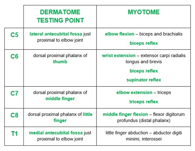
피부 분절, 근육 분절
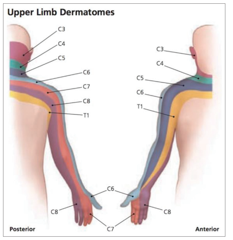
신경근 손상에서 나타나는 고유한 특징 중 하나는, 피부 분절이나 동작 기능에서 문제가 발생한다는 것이다.
왼쪽 그림은 각 신경근 손상 시 나타나는 피부 분절 통증이며, 오른쪽 그림은 각 신경근 손상 시 나타나는 동작 기능 장애를 의미한다.
예를 들면, 5번 신경근이 손상되면 어깨부터 팔 가운데 부위까지 타고 내려오는 통증이 발현되며, 팔꿈치를 굽히는 증상이나 회내시키는 동작이 제한된다는 것이다.
상완신경총의 신경근 손상에 따른 증상 정리는 아래 그림을 참고
Phrenic nerve
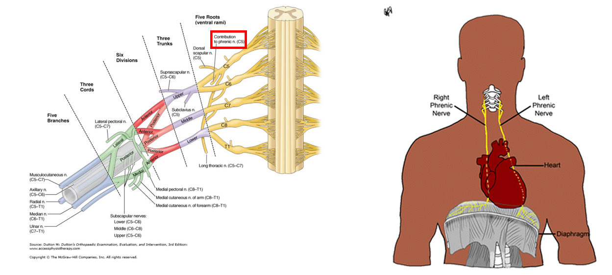
기원
C3, C4, C5 신경근에서 분지된 신경이며, 교감신경과도 연결된다.
감각 신경 지배
🔰 횡격막 힘줄 (Central tendon of the diaphragm)
🔰 흉막강(Mediastinal pleura)
운동 신경 지배
🔰 횡격막(Diaphragm)을 지배하는 유일한 운동신경
해부학
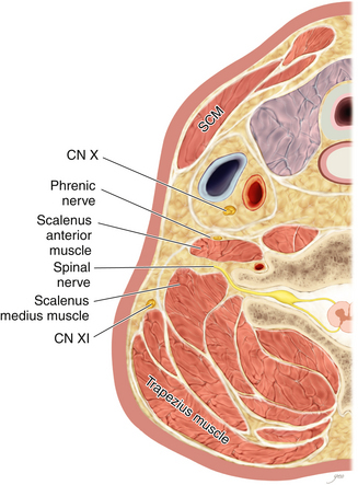
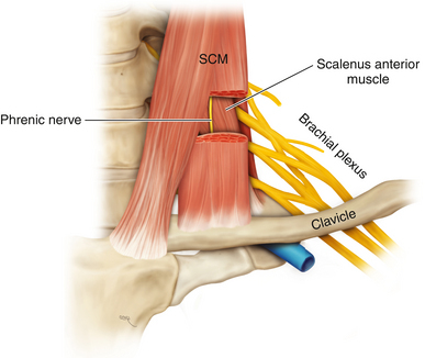
흉쇄유돌근과 전사각근 사이에 위치한 신경으로, 전사각근 바로 앞쪽에서 주행한다.
Dorsal scapular nerve

기원
C5 신경근에서 분지된 신경
감각 신경 지배
순수한 운동 신경으로, 다른 감각 부위를 지배하지 않는다.
운동 신경 지배
🔰 견갑거근 (Levator scapulae)
🔰 능형근 (Rhomboid major and minor)
해부학
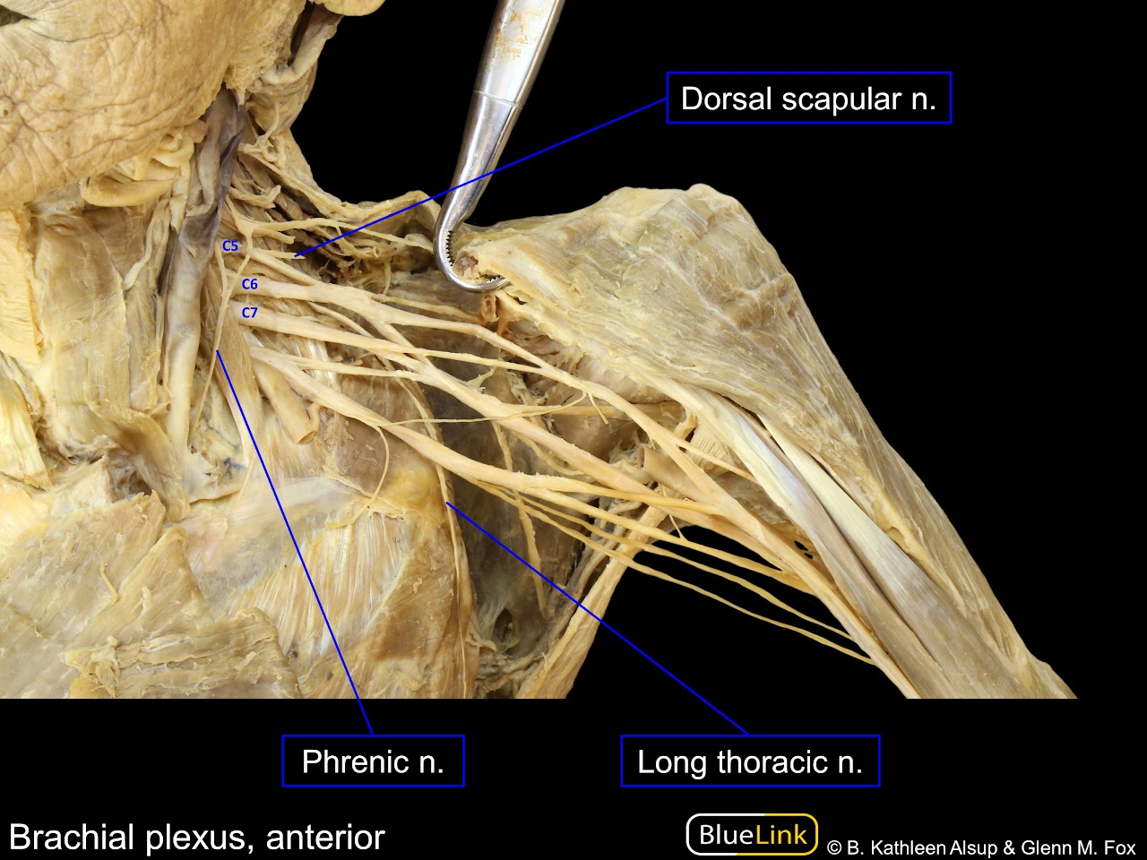
Dorsal scapular nerve는 C5신경근에서 분지되어 바로 후방으로 빠져서 날개뼈 안쪽을 타고 아래로 주행하는 걸 볼 수 있다.
Long thoracic nerve
기원
C5, C6, C7 신경근에서 분지된 신경으로, 전거근을 지배하는 유일한 운동 신경이다.
감각 신경 지배
순수한 운동 신경으로, 다른 감각 부위를 지배하지 않는다.
운동 신경 지배
🔰 전거근 (Serratus anterior)
해부학
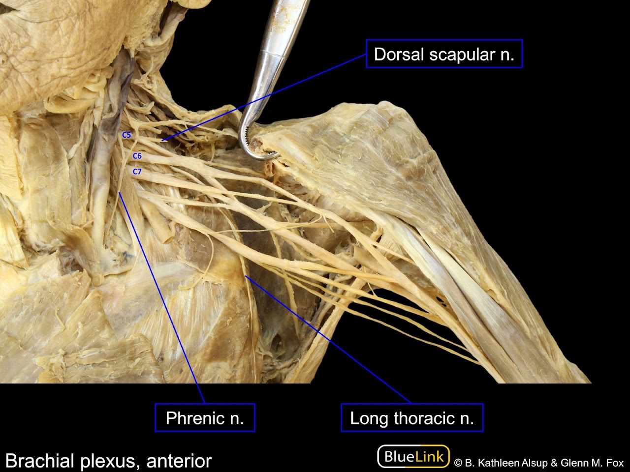
Long thoracic nerve는 C5, C6, C7신경근에서 분지되어 흉곽 바깥쪽을 타고 아래로 주행하는 모습을 볼 수 있다.
신경 간부(Trunk)
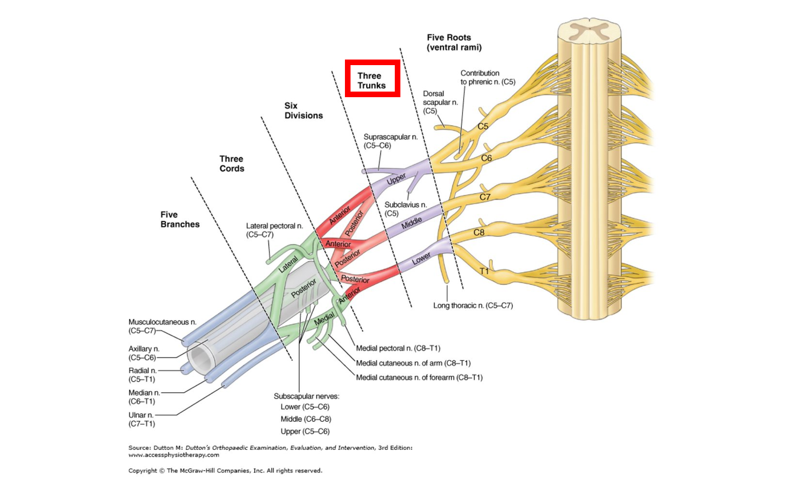
신경간부(Trunk)는 5개의 신경근에서 분지된 신경으로, C5, C6 신경근은 상부 신경간부(Upper trunk)로, C7 신경근은 중부 신경간부(Middle trunk)로, C8, T1신경근은 하부 신경간부(Iower trunk)로 합쳐진다.
Upper trunk
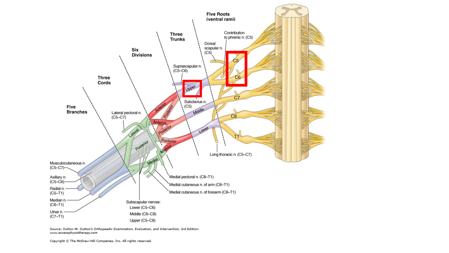
상부 신경간부는 C5, C6신경근이 합쳐진 신경으로 Suprascapular nerve(C5,C6)와 Subclavius nerve(C5)로 분지된다.
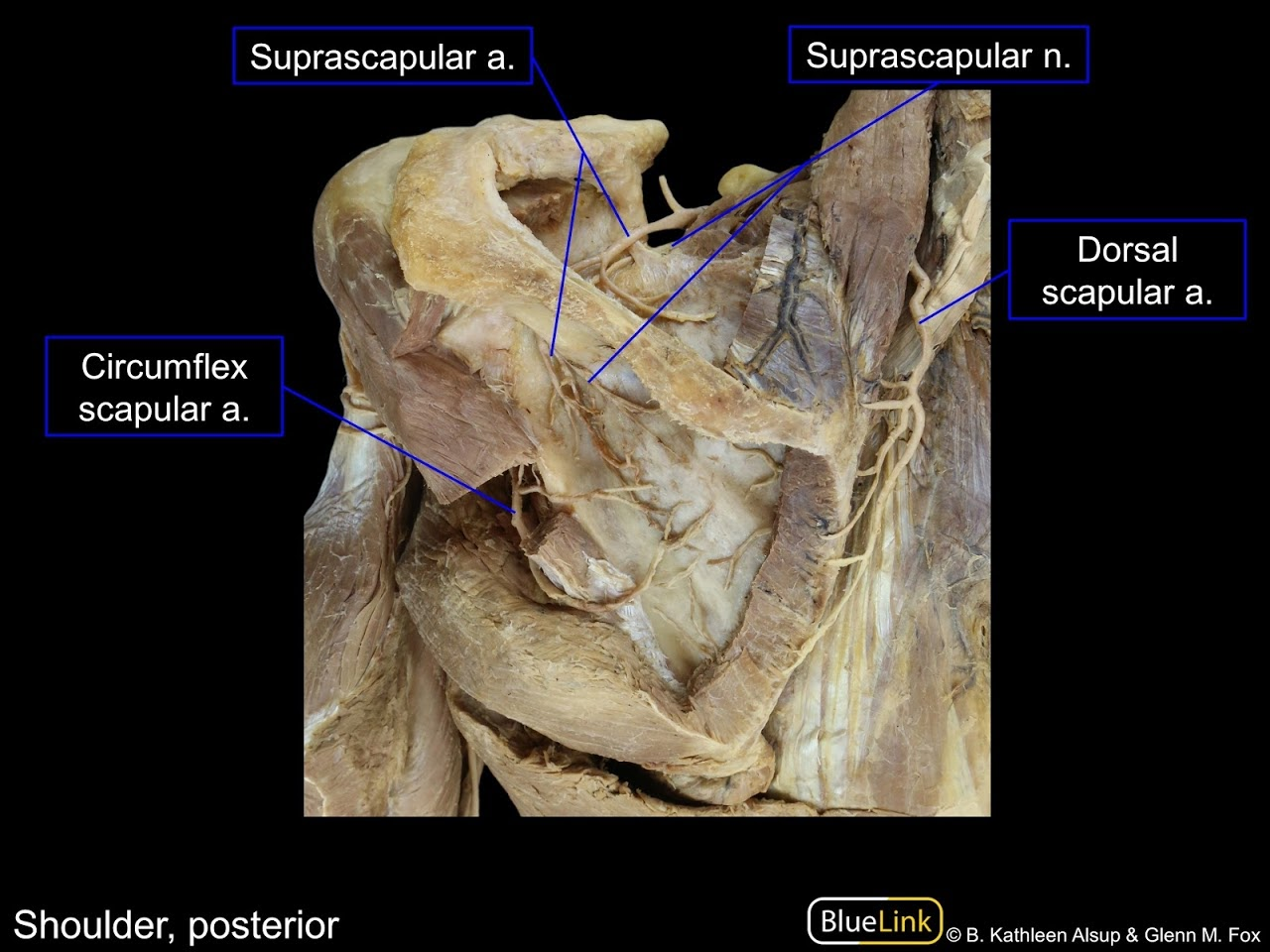
Suprascapular nerve
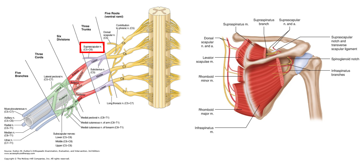
기원
C5, C6신경근에서 나온 상부 신경간부(Upper Trunk)에서 분지된 신경
감각 신경 지배
🔰 AC관절과 GH관절의 감각 신경 지배
운동 신경 지배
🔰 극상근 (Supraspinatus)
🔰 극하근 (Infraspinatus)
해부학
Suprascapular nerve는 Suprascapular notch와 Transverse scapular ligament 안쪽을 지나서 날개뼈 몸통으로 이어지는 모습을 볼 수 있다.
Subclavius nerve

기원
C5, C6신경근에서 나온 상부 신경간부(Upper Trunk)에서 분지된 신경
감각 신경 지배
순수한 운동 신경으로, 다른 감각 부위를 지배하지 않는다.
운동 신경 지배
🔰 쇄골하근 (Subclavius)
Middle trunk

중부 신경간부는 C7 신경근 으로부터 분지된 신경으로, 상부 신경간부와 다르게 별다른 신경 분지가 존재하지 않는다.
Lower trunk
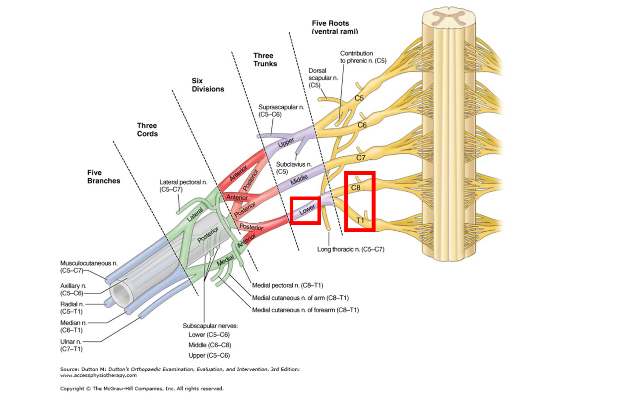
하부 신경간부는 C8, T1신경근이 합쳐진 신경으로, 상부 신경간부와 다르게 별다른 신경 분지가 존재하지 않는다.
신경 분지(Division)
신경 분지는 총 6개로 나뉘며, 굴곡근과 해당 근육들이 위치한 피부 신경을 지배하는 전방 신경 분지(Anterior division)와 신전근과 해당 근육들이 위치한 피부 신경을 지배하는 후방 신경 분지(Posterior divition)으로 구분된다.
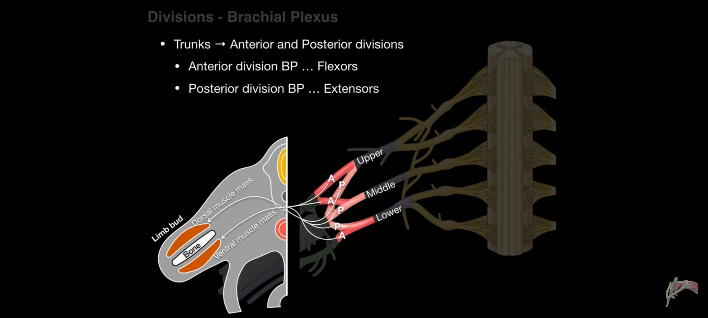
전방 분지 vs 후방 분지
신경 간부(Trunk)에서는 각각 하나의 전방 분지와 후방 분지가 나타난다.
전방 분지는 몸 앞쪽에 위치한 근육들(Vendtral muscles)을 지배하며, 후방 분지는 몸 뒤쪽에 위치한 근육들(Dorsal muscles)을 지배한다.
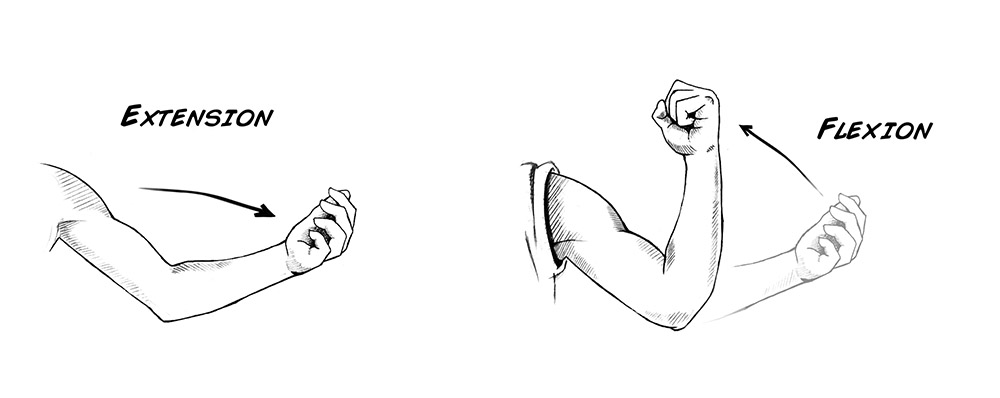
몸 앞쪽에 위치한 근육들은 대부분 팔꿈치를 굽히거나, 팔을 들어올리는 등 굴곡 기능과 연관되어 있으며 (Flexion)
몸 뒤쪽에 위치한근육들은 대부분 팔꿈치를 펴거나, 팔을 내리는 등 신전 기능과 연관되어 있다. (Extension)
신경속(Cord)
신경속은 총 3개로 나뉘며, 액와동맥(Axillary artery)를 기준으로 내측부에 위치하면 내측 신경속(Medial cord)로, 외측부에 위치하면 외측 신경속(Lateral cord), 뒤쪽에 위치하면 후방 신경속(Posterior cord)라고 부른다.
외측 코드

액와동맥을 기준으로 외측에 위치한 신경으로, 상부 신경간부와 중부 신경간부에서 분지된 전방 분지들로 구성된다.
Lateral pectoral nerve
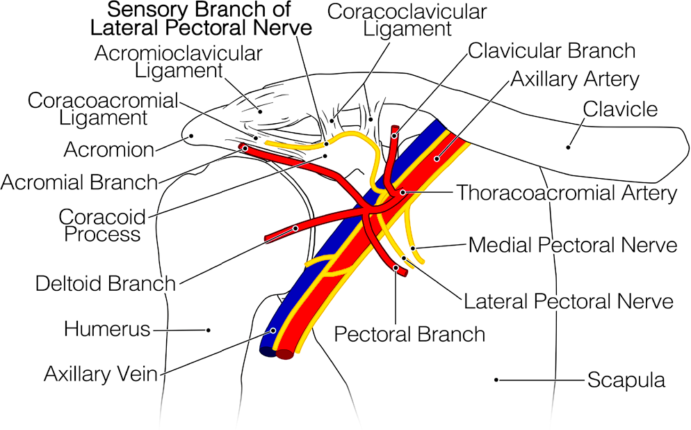
기원
C5, C6, C7 신경근에서 나온 외측 신경속(Lateral cord)에서 분지된 신경
감각 신경 지배
🔰 피부 감각을 지배하지는 않지만, 흉벽 통증을 지배하는 신경으로 가슴 수술 전 반드시 마취하는 신경이다.
운동 신경 지배
🔰 대흉근 쇄골지 (Upper clavicular part of the pectoralis major)
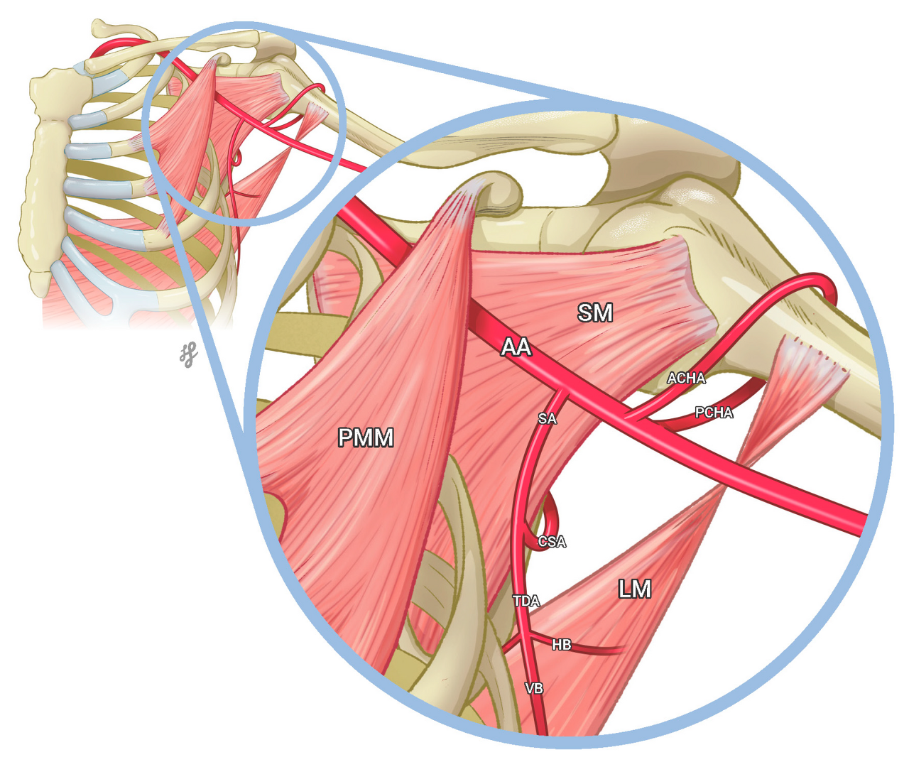
해부학
Lateral pectoral nerve가 대흉근(Pectoralis major)로 이어진 모습을 볼 수 있다.
후방 코드
액와동맥을 기준으로 후방에 위치한 신경으로, 상부 신경간부와 중부 신경간부, 하부 신경간부에서 분지된 후방 분지들로 구성된다.
Subscapular nerve
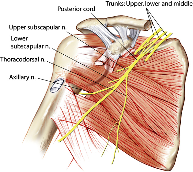
기원
C5, C6 신경근에서 나온 후방 신경속(Posterior cord)에서 분지된 신경
감각 신경 지배
순수한 운동 신경으로, 다른 감각 부위를 지배하지 않는다.
운동 신경 지배
🔰 견갑하근 (Subscapularis)
🔰 대원근 (Teres major)
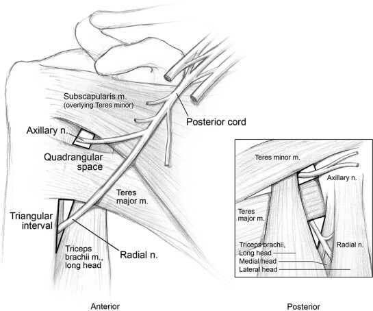
Thoracodorsal nerve
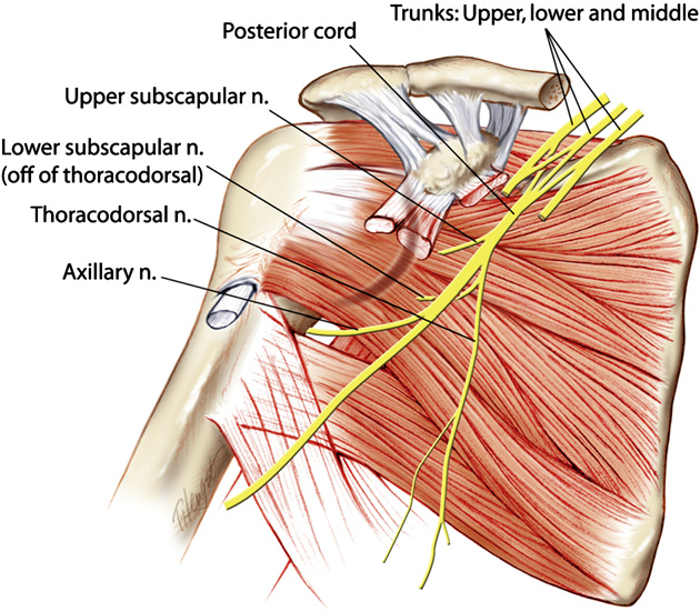
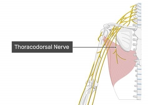
기원
C6, C7, C8 신경근에서 나온 후방 신경속(Posterior cord)에서 분지된 신경
감각 신경 지배
순수한 운동 신경으로, 다른 감각 부위를 지배하지 않는다.
운동 신경 지배
🔰 광배근 (Latissimus dorsi)
내측 코드
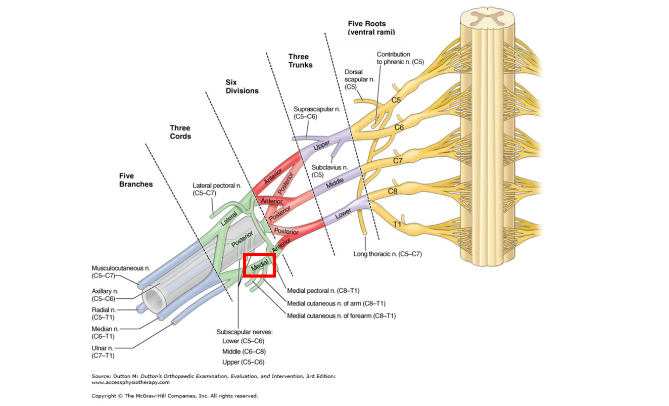
액와동맥을 기준으로 내측에 위치한 신경으로, 하부 신경간부에서 분지된 전방 분지로 구성된다.
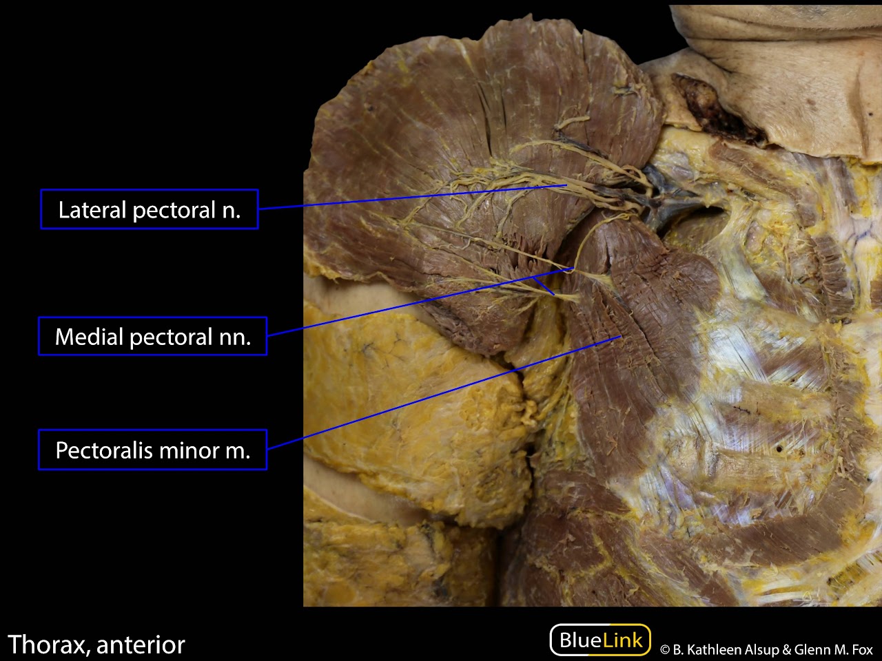
Medial pectoral nerve
기원
C8, T1 신경근에서 나온 내측 신경속(Medial cord)에서 분지된 신경
감각 신경 지배
🔰 피부 감각을 지배하지는 않지만, 흉벽 통증을 지배하는 신경으로 가슴 수술 전 반드시 마취하는 신경이다.
운동 신경 지배
🔰 소흉근 (Pectoralis minor)
🔰 대흉근 흉골지 (Lower sternocostal part of the pectoralis major)
해부학
Medial pectoral nerve가 소흉근(Pectoralis minor)에 연결된 모습을 볼 수 있다.
Medial cutaneous nerve of the forearm
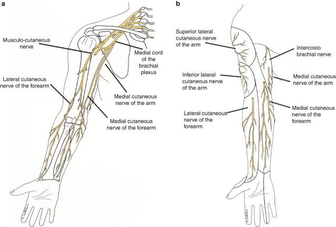
기원
C8 신경근에서 나온 내측 신경속(Medial cord)에서 분지된 신경
감각 신경 지배
🔰 이두근 부위, 전완 내측부, 팔꿈치 전면부 피부 영역
운동 신경 지배
순수한 감각 신경으로, 다른 근육 부위를 지배하지 않는다.
Medial cutaneous nerve of the arm
기원
T1 신경근에서 나온 내측 신경속(Medial cord)에서 분지된 신경
감각 신경 지배
🔰 팔 내측 3분의 1지점 까지의 피부 영역
운동 신경 지배
순수한 감각 신경으로, 다른 근육 부위를 지배하지 않는다.
신경말단 분지(Terminal branch)
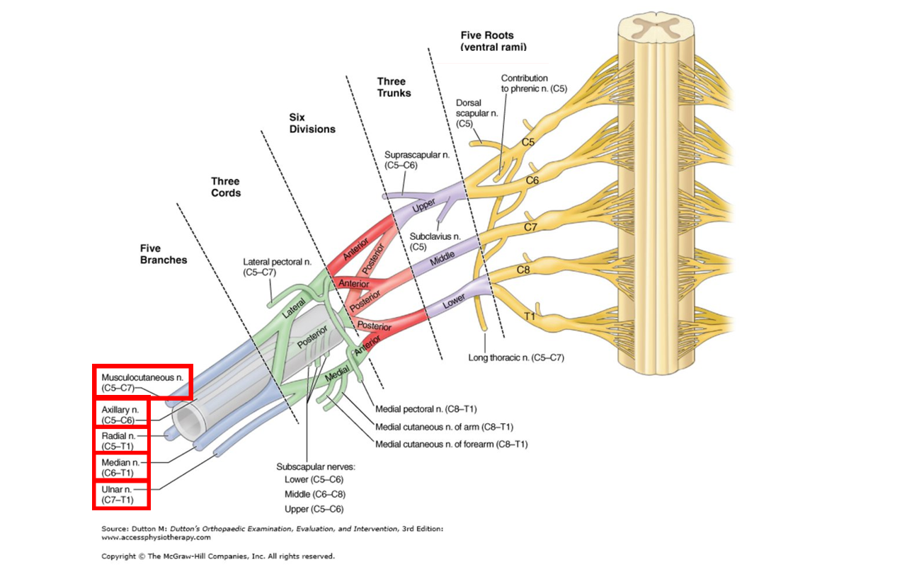
신경말단 분지는 총 5개로 분지되며, 위에서부터 근피신경(Musculocutaneous n.), 액와신경(Axillary n.), 요골신경(Radial n.), 정중신경(Median n.), 척골신경(Ulnar n.)이라 한다.
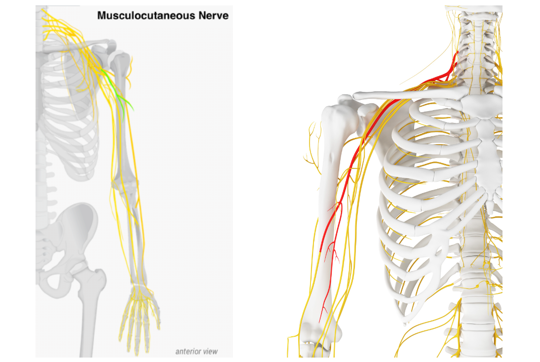
Muscaulocutaneous nerve
기원
C5, C6, C7에서 분지된 신경
감각 신경 지배
🔰 전완의 외측부 피부
운동 신경 지배
🔰 오훼완근 (Coracobrachialis)
🔰 이두근 (Biceps brachii)
🔰 상완근 (Brachialis)
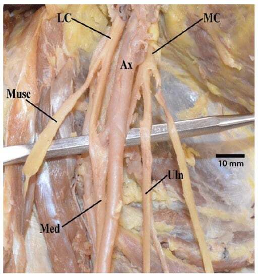
해부학

카데바를 통해 실제 근피신경의 주행경로를 이해할 수 있다.
🔰 C5, C6 Root => Superior trunk => Anterior division => Lateral cord => Musculocutaneous n.
🔰 C7 Root => Middle trunk => Anterior division => Lateral cord => Musculocutaneous n.
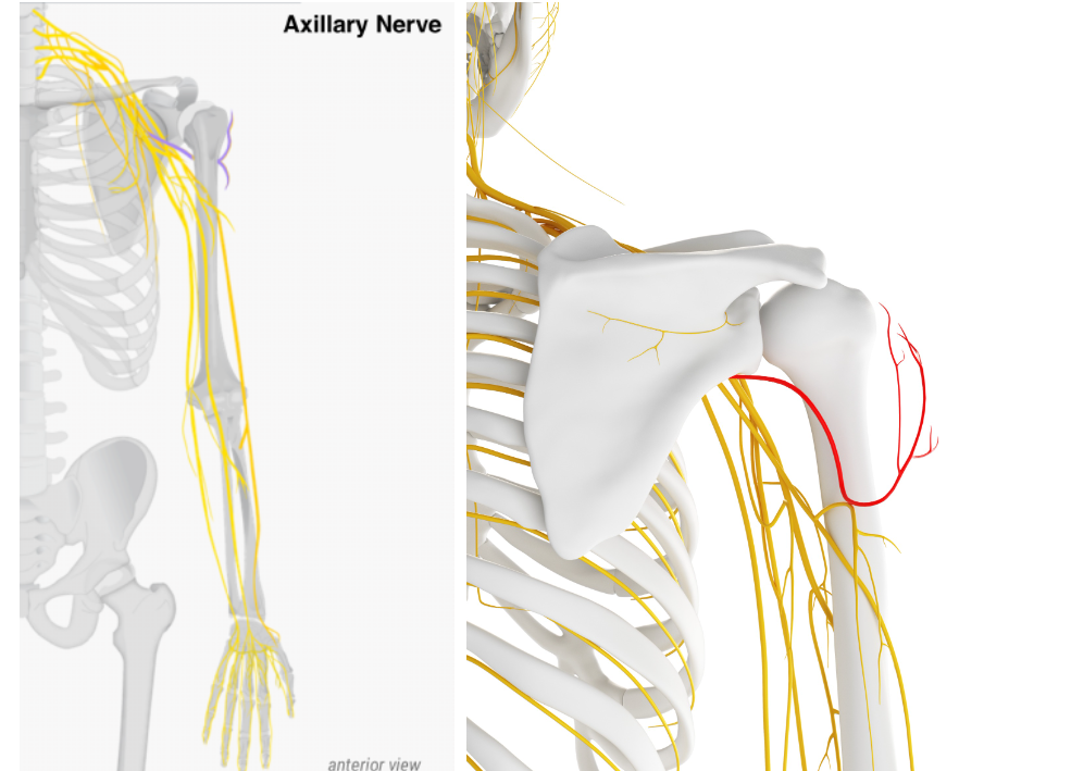
Axillary nerve
기원
C5, C6에서 분지된 신경
감각 신경 지배
🔰 어깨 외측 (Superior lateral cutaneous n.에 의해 지배됨)
운동 신경 지배
🔰 삼각근 (Deltoid)
🔰 소원근 (Teres minor)
해부학
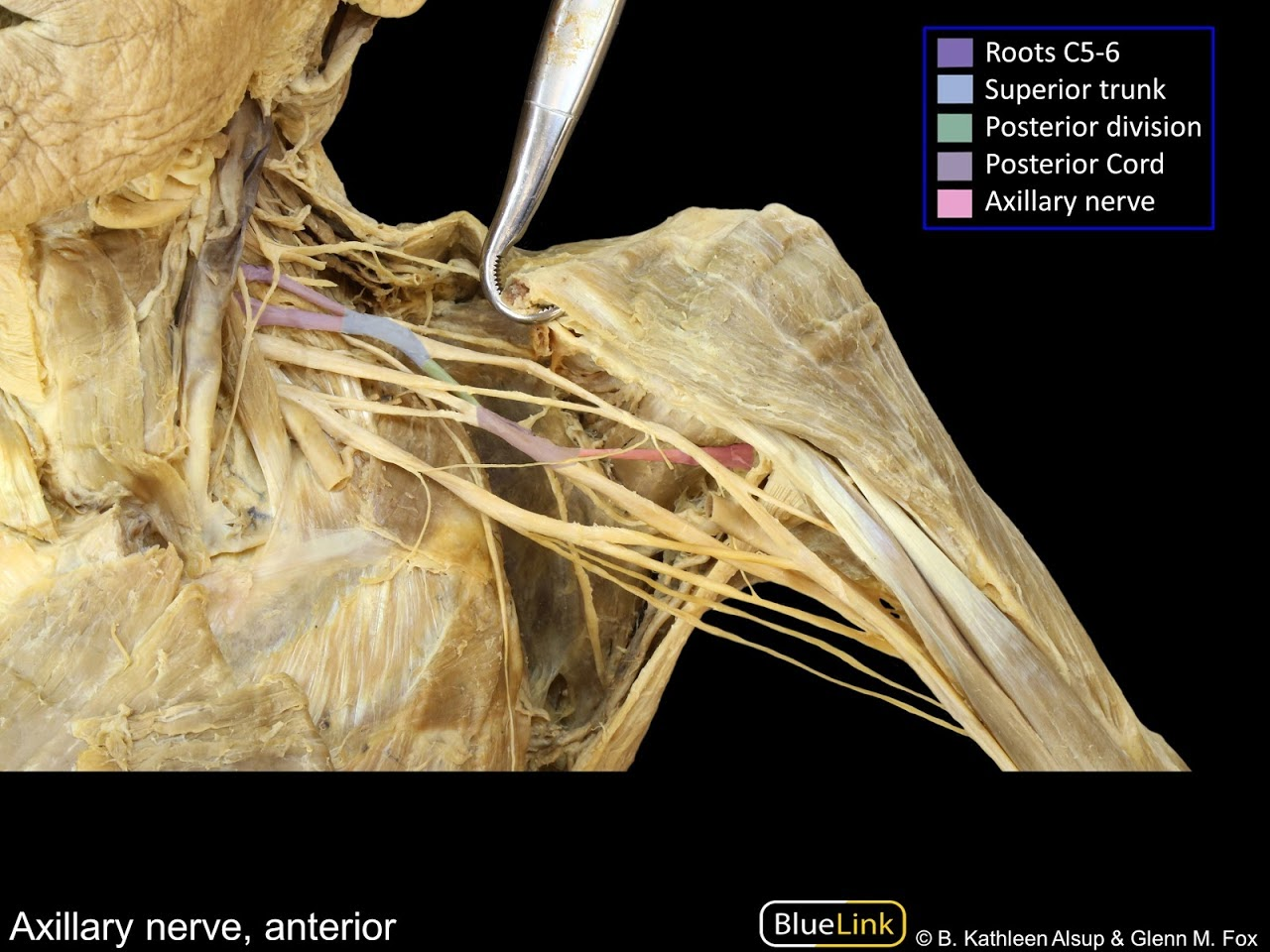
카데바를 통해 실제 액와신경의 주행경로를 이해할 수 있다.
🔰 C5, C6 Root => Superior trunk => Posterior division => Posterior cord => Axillary n.
Radial nerve
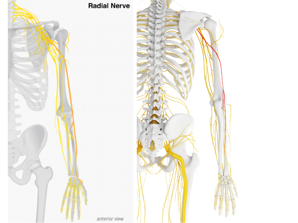
기원
C5~T1 에서 분지된 신경
감각 신경 지배
🔰 상완 외측 (Posterior cutaneous n.에 의해 지배됨)
🔰상완 뒤쪽, 전완 뒤쪽 (Inferior lateral cutaneous n.에 의해 지배됨)
🔰 1th, 2th, 3th, 4th 배측 손가락 (Dorsal digital n.에 의해 지배됨)
운동 신경 지배
🔰 외측 상완근 (Brachialis lateral part)
🔰 상완요골근 (Brachioradialis)
🔰 주근 (Anconeus)
🔰 장요측수근시근 (Extensor carpi radialis longus)
🔰 단요측수근신근 (Extensor carpi radialis brevis)
🔰 회외근 (Supinator)
해부학

카데바를 통해 실제 요골신경의 주행경로를 이해할 수 있다.
🔰 Root(C5~T1) => Trunk => Posterior division => Posterior cord => Radian n.
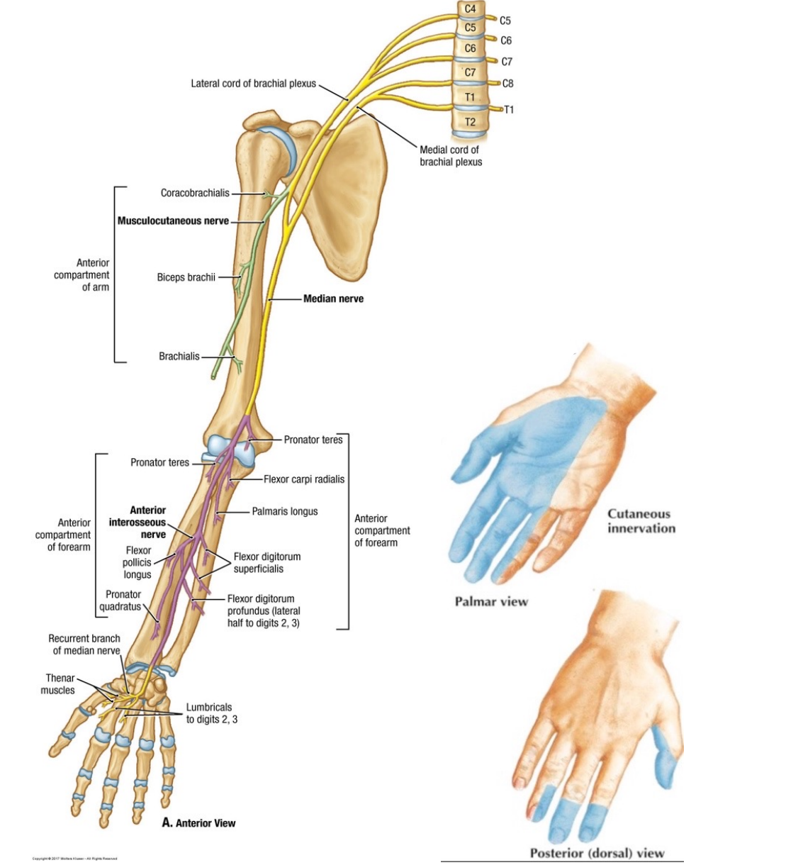
Median nerve
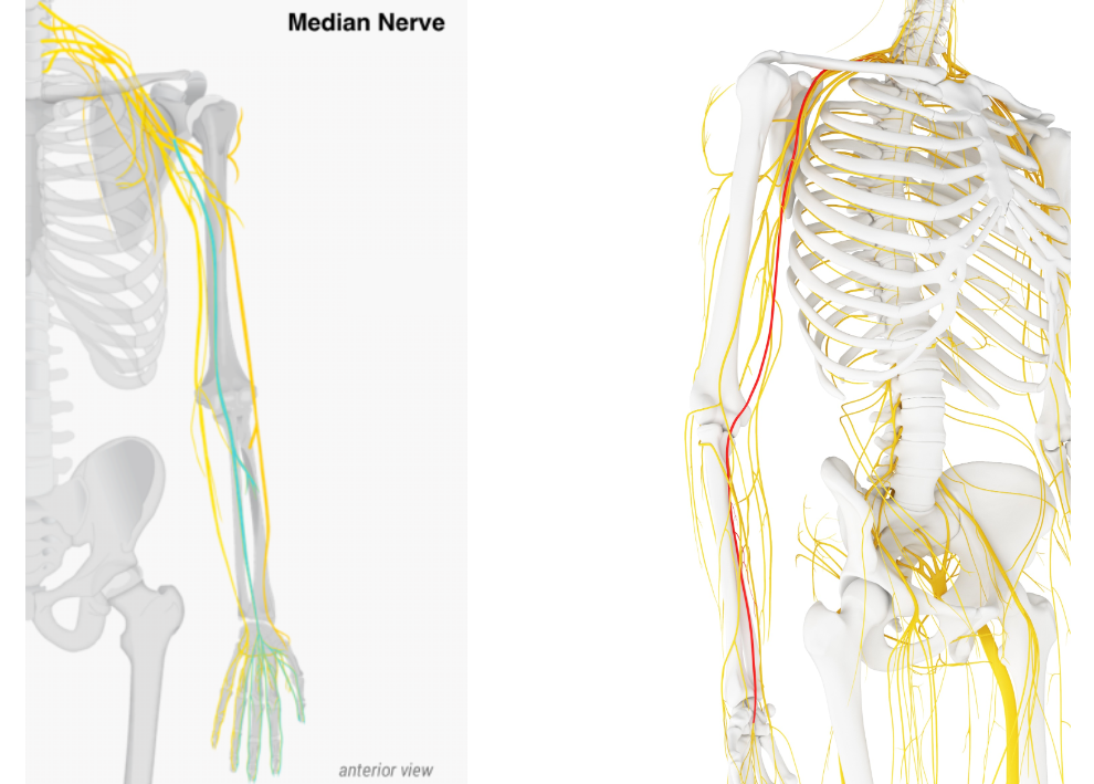
기원
C5 ~ T1에서 분지된 신경
감각 신경 지배
🔰 1th, 2th, 3th, 4th 손바닥 (Palmar cutaneus n., Proper palmar digiti n.에 의해 지배됨)
🔰 상완 뒤쪽, 전완 뒤쪽 (Inferior lateral cutaneous n.에 의해 지배됨)
운동 신경 지배
🔰 원회내근 (Pronator teres)
🔰 요측수근굴근 (Flexor carpi radialis)
🔰 장장근 (Palmaris longus)
🔰 천지굴근 (Flexor digitorum superficialis)
🔰 심지굴근 (Flexor digitorum profundus)
🔰 장무지굴근 (Flexor policis longus)
🔰 방형회내근 (Pronator quadratus)
🔰 1th, 2th 충양근 (Lumbricals manus)
🔰 단무지외전근 (Abductor policis brevis)
🔰 단무지굴근 (Flexor policis brevis)
🔰 무지대립근 (Opponens policis)
해부학

카데바를 통해 실제 정중신경의 주행경로를 이해할 수 있다.
🔰 C6 Root => Superior trunk => Anterior division => Lateral cord => Median n.
🔰 C7 Root => Middle trunk => Anterior division => Lateral cord => Median n.
🔰 C8, T1 Root => Inferior trunk => Anterior division => Medial cord => Median n.
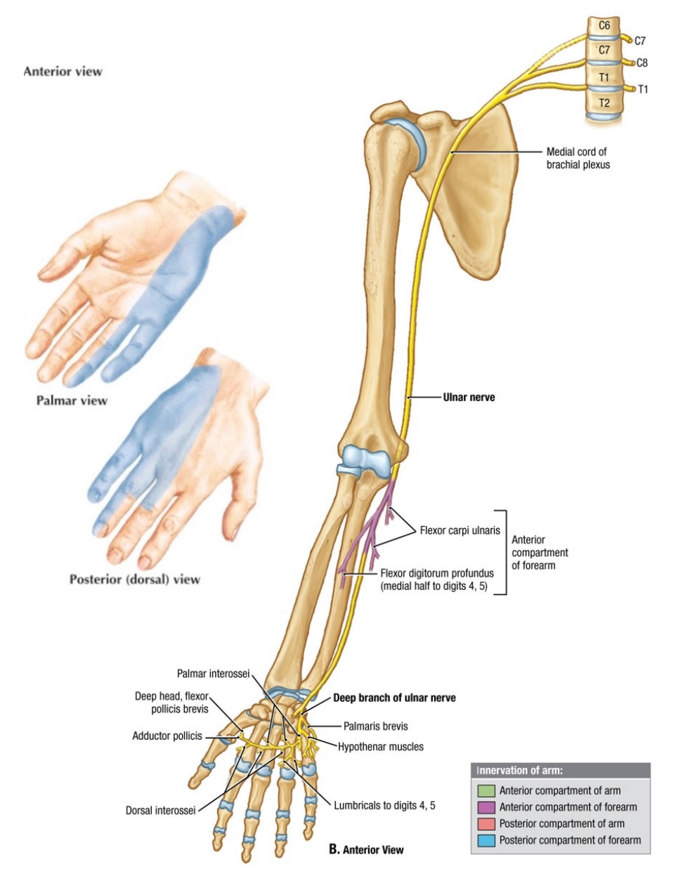
Ulnar nerve
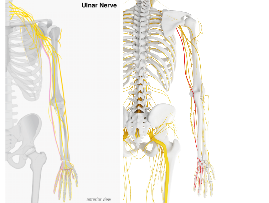
기원
C8, T1에서 분지된 신경
감각 신경 지배
🔰 4th, 5th 손바닥 (Palmar cutaneous n.에 의해 지배됨)
🔰 4th, 5th 손등 (Dorsal cutaneous n.에 의해 지배됨)
운동 신경 지배
🔰 척측수근굴근 (Flexor carpi ulnaris)
🔰 내측 심지굴근 (Medial half of flexor digitorum profundus)
🔰 단장근 (Palmaris brevis)
🔰 소지외전근 (Abductor digiti minimi)
🔰 단소지굴근 (Flexor digiti minimi brevis)
🔰 소지대립근 (Opponens digiti minimi)
🔰 3th, 4th 충양근 (Lumbricals)
🔰 골간근 (Interossei)
🔰 무지내전근 (Adductor pollicis)
해부학

카데바를 통해 실제 척골신경의 주행경로를 이해할 수 있다.
🔰 C8, T1 Root => Inferior trunk => Anterior division => Medial cord => Ulnar n.
상완신경총의 모식도
신경 분지 경로

그 외 참고하면 좋은 자료
참고
Grahn, P. (2021). Improving shoulder function in brachial plexus birth injury (Doctoral dissertation, Väitöskirja. Helsinki: Helsingin yliopisto, lääketieteellinen tiedekunta).
Walji, A. H., & Tsui, B. C. (2016). Clinical anatomy of the brachial plexus. Pediatric Atlas of Ultrasound-and Nerve Stimulation-Guided Regional Anesthesia, 149-163.
Llusá, M., Morro, M. R., Casañas, J., & Moore, A. M. (2021). Surgical anatomy of the brachial plexus. In Operative Brachial Plexus Surgery: Clinical Evaluation and Management Strategies (pp. 19-39). Cham: Springer International Publishing.
Schünke, M., Schulte, E., Schumacher, U., Ross, L. M., & Lamperti, E. D. (2014). Thieme atlas of anatomy: general anatomy and musculoskeletal system. Thieme Medical Publishers, Incorporated.
Sumarwoto, T., Suroto, H., Mahyudin, F., Utomo, D. N., Hadinoto, S. A., Abdulhamid, M., ... & Rhatomy, S. (2021). Brachial plexus injury: recent diagnosis and management. Open Access Macedonian Journal of Medical Sciences, 9, 13-24.
Dutton, M. (2012). Dutton's Orthopaedic examination, evaluation, and intervention. (No Title).
Bentley, J. N., & Yang, L. J. S. (2015). Anatomy of the Posterior Cord and Its Branches. In Nerves and Nerve Injuries (pp. 563-574). Academic Press.
Branches of the Brachial Plexus" by University of British Columbia Clinical Anatomy
Oliver, K. A., & Ashurst, J. V. (2018). Anatomy, thorax, phrenic nerves.
Goff, R. P., Spencer, J. H., & Iaizzo, P. A. (2016). MRI reconstructions of human phrenic nerve anatomy and computational modeling of cryoballoon ablative therapy. Annals of biomedical engineering, 44, 1097-1106.
Bishop, K. N., & Varacallo, M. (2017). Anatomy, shoulder and upper limb, dorsal scapular nerve.
Hida, K. (2019). Dorsal Scapular Nerve Block (Landmark Method). Nerve Blockade and Interventional Therapy, 101-103.
Olamikan, S., & Karl, H. W. (2016). Long thoracic nerve entrapment. Peripheral nerve entrapments: clinical diagnosis and management, 291-303.
Basta, M., Sanganeria, T., & Varacallo, M. (2020). Anatomy, shoulder and upper limb, suprascapular nerve.
Finneran IV, J. J. (2022). Upper Extremity Blocks: Suprascapular Nerve Block. In Anesthesiology In-Training Exam Review: Regional Anesthesia and Chronic Pain (pp. 135-139). Cham: Springer International Publishing.
Leurcharusmee, P., Maikong, N., Kantakam, P., Navic, P., Mahakkanukrauh, P., & Tran, D. Q. (2021). Innervation of the clavicle: a cadaveric investigation. Regional Anesthesia & Pain Medicine, 46(12), 1076-1079.
Patel, N. T., & Smith, H. F. (2023). Clinically Relevant Anatomical Variations in the Brachial Plexus. Diagnostics, 13(5), 830.
Chu, B., & Bordoni, B. (2022). Anatomy, Thorax, Thoracodorsal Nerves. In StatPearls [Internet]. StatPearls Publishing.
Jeno, S. H., & Varacallo, M. (2023). Anatomy, back, latissimus dorsi. In StatPearls [Internet]. StatPearls Publishing.
Celli, A., & Celli, L. (2022). Cutaneous and Muscular Nerves. In Atlas of Elbow Surgery (pp. 35 - 47). Cham: Springer International Publishing.
Kasper, J. C., Itamura, J. M., Tibone, J. E., Levin, S. L., & Stevanovic, M. V. (2008). Human cadaveric study of subscapularis muscle innervation and guidelines to prevent denervation. Journal of shoulder and elbow surgery, 17(4), 659-662.
Eckmann, M. S., Lai, B. K., Uribe III, M. A., Patel, S., & Benfield, J. A. (2019). Thermal radiofrequency ablation of the articular branch of the lateral pectoral nerve: a case report and novel technique. A&A Practice, 13(11), 415-419.
Desai, S. S., & Varacallo, M. (2018). Anatomy, shoulder and upper limb, musculocutaneous nerve.
https://sites.google.com/a/umich.edu/bluelink/curricula/first-year-medical-curriculum/sequence-7-neuroanatomy/shoulder-and-brachial-plexus/shoulder-and-brachial-plexus-written-learning-objectives
Essentials of Regional Anesthesia Anatomy. (n.d.). [Untitled Mytomtomes] Retrieved October 23, 2015, from http://www.nysora.com/mobile/regional-anesthesia/3012-essentials-of-regional-anesthesia-anatomy.html
https://www.youtube.com/watch?v=RLJ8aUw468M
https://med.libretexts.org/Bookshelves/Anatomy_and_Physiology/Human_Anatomy_%28Lange_et_al.%29/12%3A_Peripheral_Nervous_System/12.04%3A_Spinal_Nerves_and_Their_Branches
https://antranik.org/peripheral-nervous-system-spinal-nerves-and-plexuses/
https://www.osmosis.org/learn/Anatomy_of_the_brachial_plexus
https://emedicine.medscape.com/article/1877731-overview
'Exercise is medicine > 기능 해부학' 카테고리의 다른 글
| 발목 인대의 해석 [측면 인대] (0) | 2023.08.06 |
|---|---|
| 요천추신경총 해부학 (0) | 2018.03.19 |
| 견갑대 해부학 (2) | 2018.03.16 |
| 골반 해부학 (1) | 2018.03.15 |
| 목 뼈 해부학 (0) | 2018.03.12 |


- Total
- Today
- Yesterday